Musculoskeletal Ultrasound Used in the Diagnosis of Carpal Tunnel Syndrome
Musculoskeletal Ultrasound Used in the Diagnosis of Carpal Tunnel Syndrome
Musculoskeletal Ultrasound Used in the Diagnosis of Carpal Tunnel Syndrome

Over the past 20 years, neuromuscular ultrasound has been introduced into electrodiagnostic laboratories as a complement to nerve conduction studies and electromyography for the diagnosis of a variety of nerve and muscle conditions.
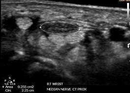
Carpal Tunnel Syndrome (CTS) is the most commonly studied condition with neuromuscular ultrasound, and individuals with CTS have displayed ultrasonographic evidence of focal enlargement of the median nerve at the wrist.
But the most important use of, neuromuscular ultrasound for median nerve entrapment at the wrist is to identify the causes of the median mononeuropathy and structural anomalies that could not be detected with electrodiagnostic studies alone, such as compressive cysts, tumors, bifid median nerves and vessels.
A recent study published at Muscle & Nerve demonstrated that in at least 20% of the CTS cases ultrasonography of the median nerve can identify one of the above causes as the culprit for the condition and therefore intervention can be more appropriate and effective.